Mirror users: Please use the PhysioNet master server as needed if files of interest within this database are inaccessible from a mirror.
This is version 1.0 of the MIMIC-III Waveform Database (August 2017; 67,830 records.)
This database is described in
Please cite this publication when referencing this material, and also include the standard citation for PhysioNet:
The MIMIC-III Waveform Database contains thousands of recordings of multiple physiologic signals ("waveforms") and time series of vital signs ("numerics") collected from bedside patient monitors in adult and neonatal intensive care units (ICUs). It is a companion to the MIMIC-III Clinical Database, which contains detailed clinical information for many of the patients represented in the Waveform Database. The MIMIC-III Waveform Database Matched Subset contains 22,317 waveform records and 22,247 numerics records, which have been matched and time-aligned with 10,282 MIMIC-III Clinical Database records.
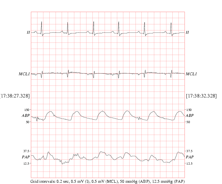
Recorded waveforms and numerics vary depending on choices made by the ICU staff. Waveforms almost always include one or more ECG signals, and often include continuous arterial blood pressure (ABP) waveforms, fingertip photoplethysmogram (PPG) signals, and respiration, with additional waveforms (up to 8 simultaneously) as available. Numerics typically include heart and respiration rates, SpO2, and systolic, mean, and diastolic blood pressure, together with others as available. Recording lengths also vary; most are a few days in duration, but some are shorter and others are several weeks long.
Use the PhysioBank ATM to view any desired record in this database, to export it in a variety of formats, or to perform a variety of other operations on it.
What's New
This database is closely related to the older MIMIC II Waveform Database, which (as of version 3.2) shares the same underlying set of records. The only difference between the two databases lies in the Matched Subset, which has been matched with the MIMIC-III Clinical Database (version 1.4.)
Version History
For more information about past releases of the MIMIC II Waveform Database, see its version history section.
| Version | Released | Records |
|---|---|---|
| 1.0 | August 2017 | 67,830 |
Organization of the Database
Each recording comprises two records (a waveform record and a matching numerics record) in a single record directory ("folder") with the name of the record. To reduce access time, the record directories have been distributed among ten intermediate-level directories (listed below). The names of these intermediate directories (30, 31, ..., 39) match the first two digits of the record directories they contain.
In almost all cases, the waveform records comprise multiple segments, each of which can be read as a separate record. Each segment contains an uninterrupted recording of a set of simultaneously observed signals, and the signal gains do not change at any time during the segment. Whenever the ICU staff changed the signals being monitored or adjusted the amplitude of a signal being monitored, this event was recorded in the raw data dump, and a new segment begins at that time.
Each composite waveform record includes a list of the segments that comprise it in its master header file. The list begins on the second line of the master header with a layout header file that specifies all of the signals that are observed in any segment belonging to the record. Each segment has its own header file and (except for the layout header) a matching (binary) signal (.dat) file. Occasionally, the monitor may be disconnected entirely for a short time; these intervals are recorded as gaps in the master header file, but there are no header or signal files corresponding to gaps.
The numerics records (designated by the letter n appended to the record name) are not divided into segments, since the storage savings that would be achieved by doing so would be relatively little.
Physiologic waveform records in this database contain up to eight simultaneously recorded signals digitized at 125 Hz with 8-, 10-, or (occasionally) 12-bit resolution. Numerics records typically contain 10 or more time series of vital signs sampled once per second or once per minute.
An example will make this arrangement clear:
- Intermediate directory 31 contains all records with names that begin with 31.
- Record directory 3141595 is contained within intermediate directory 31.
- All files associated with physiologic waveform record 3141595 and its
companion numerics record 3141595n are contained within record directory
31/3141595.
- The first line of the master header file for waveform record 314595 (31/3141595/3141595.hea) indicates that the record is 242353557 sample intervals (about 22 days at 125 samples per second) in duration, and that it contains 427 segments and gaps. (See header(5) in the WFDB Applications Guide for details on the format of this text file.) The first segment is named 3141595_0001, and it is 2888500 sample intervals (6 hours, 15 minutes, and 8 seconds, at 125 samples per second) in duration. At the end of the master header file, a comment (# Location: nicu) specifies the ICU in which the recording was made (the neonatal ICU in this case).
- The layout header file for this record (31/3141595/3141595_layout.hea) indicates that five ECG signals (I, II, III, AVR, and "V"), a respiration signal, and a PPG signal are available during portions of the record. (The five ECG signals are not all available simultaneously.)
- The header file for the first segment of this record (31/3141595/3141595_0001.hea) shows that a PPG signal ("PLETH"), a respiration signal, and ECG leads II and AVR are available throughout this initial segment.
- The matching numerics record is named 3141595n, and its header file (31/3141595/3141595n.hea) shows that it is 1938730 sample intervals (about 22 days at 1 sample per second) in duration, and that it contains heart rate (HR, from ECG, as well as PULSE, from one or more pulsatile signals), noninvasive blood pressure (raw as well as systolic, diastolic, and mean), respiration rate, and SpO2.
Any WFDB application can read any waveform record from this database directly from the PhysioNet web server (i.e., without downloading the record first) using a record name of the form mimic3wdb/3x/3xyyyyy/. Numerics records can be read using the longer form mimic3wdb/3x/3xyyyyy/3xyyyyyn (note that the final 3xyyyyy must be repeated and followed by n to specify the numerics record).
For example, if you have installed the WFDB Software Package, you can read the first 10 seconds of waveform record 3141595 using this rdsamp command:
rdsamp -r mimic3wdb/31/3141595/ -p -v -t 10
To read the first 10 seconds of the matching numerics record 3141595n, use this command instead:
rdsamp -r mimic3wdb/31/3141595/3141595n -p -v -t 10
Notice that the first command produces 1250 samples of each waveform (125 samples per second, for 10 seconds), but the second command produces only 10 samples of each vital sign (1 sample per second, for 10 seconds). See How to obtain PhysioBank data in text form for details about using rdsamp.
Clinical Correlates
The MIMIC-III Clinical Database contains detailed clinical information about most of the subjects represented in the MIMIC-III Waveform Database. Since the contents of each database were collected independently, in partially deidentified form, matching the clinical data with the waveform data is a non-trivial task, and only a subset of MIMIC-III Waveform Database records, located in the MIMIC-III Waveform Database Matched Subset, has been matched with MIMIC-III Clinical Database records.
In these cases, the matches provide additional information about the subjects, including age, gender, and detailed clinical information collected during (and in some cases before and after) the periods that have been recorded in the Waveform Database records. For more information, apply for access to the MIMIC-III Clinical Database (a data use agreement is required).
Multiple recordings of a given patient, which may exist (for example) if that patient was admitted more than once to any of the study ICUs during the study period, do not have related MIMIC-III Waveform Database record names; it will be necessary to refer to the Matched Subset to discover any such cases.
Technical Limitations
Waveforms or numerics missing: Occasionally, technical limitations of the data acquisition system make it possible to create a physiologic waveform record but not a numerics record, or vice versa.
A given signal may not be available throughout an entire record. Records in the MIMIC-III Waveform Database vary in length; some are several weeks in duration. It is common for the physiologic signals to be interrupted or changed occasionally during recordings of such long duration. When using a viewer such as the PhysioBank ATM, all signals available at any time during a record are listed, although in most cases only a subset is visible at any given time.
Gaps and patient identification. The waveform and numerics records have been extracted from raw data dumps collected from the bedside monitors using a facility provided by the monitor manufacturer. The raw data dumps contain files of data collected from a single patient monitor during a single monitoring session (which may last days or weeks). Usually the monitoring session ends when the patient is discharged, so that the data in a single file come from a single patient. Occasionally, however, the monitor is not reset when the patient is discharged, and the session continues after a new patient has been admitted; in this case the raw data file contains data from two (or more) patients, with a gap (an interval during which no waveforms or numerics are recorded) that is typically an hour or more in duration. Such gaps may also appear if the monitor is temporarily disconnected (for example, for a laboratory test) and then reconnected to the same patient. Since the raw data files do not usually contain patient identifiers, it is not trivial to determine with certainty if the data before and after a gap were collected from the same patient.
Ideally, each MIMIC-III Waveform Database record should contain data from only one patient. All raw data files containing gaps of an hour or more have been split into separate records in order to decrease the likelihood that any record contains data from multiple patients. An ongoing project is to examine the sets of records created this way, matching them with MIMIC-III Clinical Database records when possible, to determine if and how they should be reassembled.
Inter-waveform alignment problems: The method used for MIMIC waveform data extraction was not designed for inter-waveform analysis. The waveform data contain unspecified/unknown filtering delays and/or unknown inter-channel delays, which may not be constant in a given record. Therefore, although the ECGs are time-aligned with each other, there may be a (changing) delay of up to 500ms between any of the other waveforms in the data. For example, the pulse transit time measured between different waveforms may be unreliable (either in absolute or relative terms).
ECG limitations: The ECG signals in the waveform records were originally sampled with 12-bit precision at a high sampling rate, and were then scaled and decimated to 500 samples per second (per signal). The scaling reduced the effective amplitude resolution from 12 bits to 9 or 10 bits in typical cases, and as little as 7 bits in some cases. From each set of 4 consecutive decimated samples of the same ECG signal, one was recorded (chosen using a turning-point compressor, a technique sometimes called "peak-picking"). The result is an ECG signal sampled 125 times per second, but at intervals that vary between 2 and 14 ms (averaging 8 ms). Since the interval between any given pair of samples was not available to us, the reconstructions of the ECG signals assume uniform 8 ms intervals. These signals with reduced time and amplitude resolution, and sampling jitter introduced by the "peak-picking", were the only ECG signals that were possible to capture from the ICU monitors. Although ECGs reconstructed in this way can be readily interpreted visually, they are unsuitable as input for certain algorithms for ECG analysis, particularly those that are sensitive to frequency-domain features of the signal. Note that these limitations apply only to the ECG signals, not to the other signals, which were originally sampled at uniform 8 ms intervals (125 samples per second) and were not scaled prior to capture.
Name Last modified Size Description
Parent Directory -
mimic3wdb.sh 2018-09-07 16:00 2.7K
matched/ 2017-08-07 18:17 -
DOI 2017-08-07 18:15 20
RECORDS 2017-08-04 17:28 795K list of record names
|
If you would like help understanding, using, or downloading content, please see our Frequently Asked Questions. If you have any comments, feedback, or particular questions regarding this page, please send them to the webmaster. Comments and issues can also be raised on PhysioNet's GitHub page. Updated Friday, 28 October 2016 at 16:58 EDT |
PhysioNet is supported by the National Institute of General Medical Sciences (NIGMS) and the National Institute of Biomedical Imaging and Bioengineering (NIBIB) under NIH grant number 2R01GM104987-09.
|